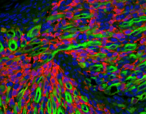
Nanotechnology has contributed to the advances in our ability to see different things at the nanoscale. Microscopy has advanced from the very early days of microscopes being a single glass lens to very advanced instruments with nanometer resolution. We can see lots of stuff with high resolution and even in 3D. And sometimes the picture is just neat. Scientists at St. Judes Hospital used a confocal microscope to study the progression of a soft tissue cancer to understand the origins of these cancer cells. It was once believed these cancer cells came from muscle tissue but in fact are from cells that make up blood vessels. The technique and the image involve immunostaining, using antibodies against bind to different things. The antibodies are ‘painted’ with a dye that results in different colors. The cool image may help doctors understand
 how an important disease like cancer develops and how it might be cured.